FeaturedYPE html> Zhang Lab | Neuroscience at VCU
Virginia Commonwealth University

Understanding the Neural Basis of Social Behavior

Connecting with others is important for our health and well-being. But in conditions such as autism, depression, and Alzheimer's disease, social withdrawal can make connection difficult, even when social support could help.

We study how social connection works at three levels. In human populations, large datasets show what social connection does to health. In freely interacting animals, we record and manipulate the circuits at work while the interaction unfolds. And in embodied digital twins, complete nervous systems modelled inside a moving body, we can follow activity through every neuron at once. No single level explains social connection on its own. Read together, they point toward why it breaks down, and how it might be restored.

What We Study

One question at three levels: what social connection does to health, how the brain produces it, and what a complete nervous system can reveal when nothing is hidden.

We study social behaviors of human populations because:

Only in people can we see what social connection actually does to health, at the scale of whole populations. Large human datasets let us ask how the social environment and everyday social behavior relate to physical and mental health: who becomes isolated, and what that isolation costs. The relationship runs in both directions: poor health narrows social life, and a narrowed social life worsens health. These datasets establish that the link is real and how much it matters. What they cannot show is what the brain is doing, which is where animals come in.
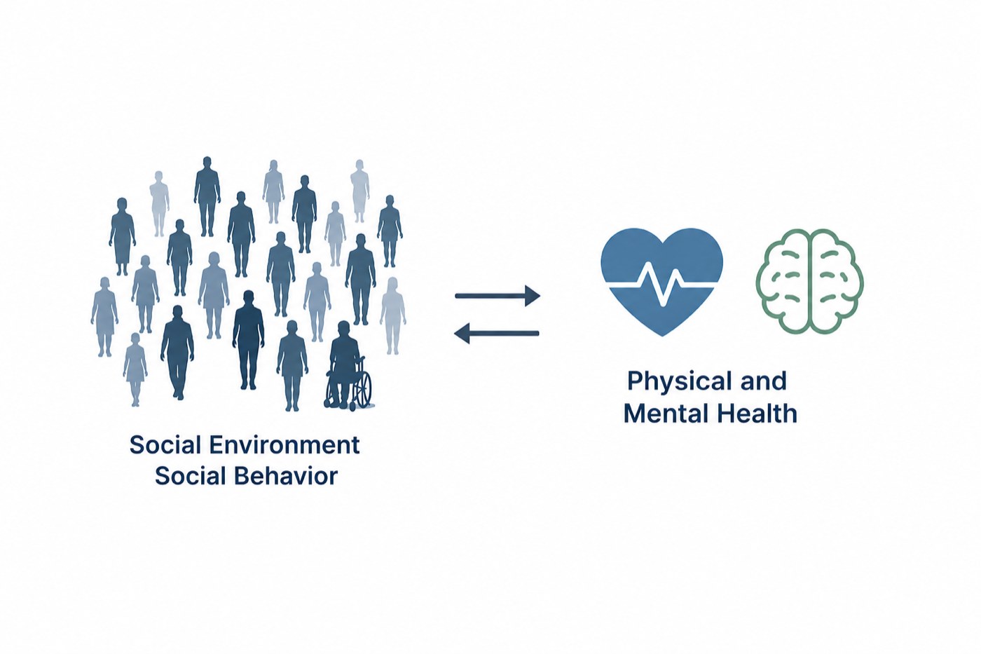
Social environment and social behavior linked by two-way arrows to physical and mental health
Social environment and social behavior are reciprocally linked to physical and mental health across populations.

We study social behaviors of freely interacting animals because:

Animals such as mice have a rich social life of their own, which is what lets us turn these questions into mechanistic ones we can test directly in the brain. In this video, a bystander mouse works to help a companion in distress, pulling its tongue out of the mouth, which helps keep the airway open, and these efforts often allowed the unresponsive mouse to recover. Strikingly, mice do this for individuals they already know, but not for strangers. Because the behavior is this specific, we can record and manipulate the circuits that produce it while the interaction is unfolding. What we still cannot do in a living animal is watch every neuron at once.

A bystander mouse pulls the tongue of an unconscious familiar mouse out of the mouth, helping keep the airway open. Read our Science paper.

We study social behaviors of embodied digital twins of the biological nervous system because:

Even the best recording reaches only a fraction of the neurons in a brain, and it cannot tell us how all of them are wired. Reconstructed nervous systems close that gap. Two animals are now mapped completely, body and nervous system together: the fly and C. elegans. With every neuron and connection known, we can simulate activity across the entire nervous system and watch how it drives movement and social behavior. Nothing is hidden in a model like this: we can follow a signal from one identified neuron all the way to the behavior it produces, and we can watch activity in both partners at once through an entire social interaction, which no experiment can currently do. Circuit hypotheses that are out of reach in a living animal can be tested here first, and the promising ones taken back to the bench.

A fully reconstructed Drosophila central nervous system and body. Cell bodies carry simulated neuronal activity, with a single identified descending neuron shown in anatomical context alongside the whole-body movement it drives.
Two digital twins interacting. Each fly's reconstructed cells carry their own simulated activity, shown above as independent signals, while their movements and gestures play out below. This is the point of an embodied twin: we can watch neuronal activity in both partners at the same time, throughout a social interaction.
A completely reconstructed C. elegans, body and nervous system together. Every one of its neurons and their connections are known, so the whole animal can be modelled at once: the nerve ring, ventral cord, and commissures are visible through the body as it moves.

How We Do It

The techniques and projects we build to get from social behavior to the circuits behind it.

AI-Powered Analysis

Automatic Social Behavior Analysis

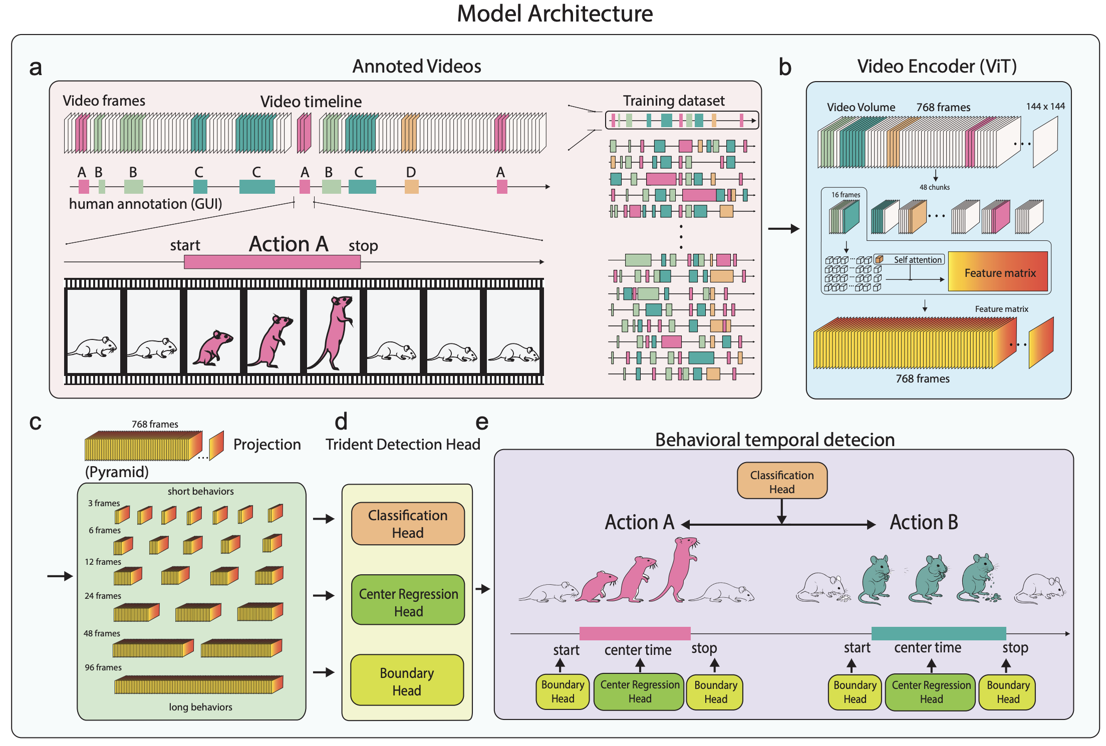
Natural social interactions unfold over long periods of time. To understand them, we need to track behavior continuously. Manual annotation is slow, tedious, and subjective. We therefore developed automatic approaches that can analyze social behavior accurately and at scale.

Our machine learning system tracks individual animals and automatically identifies what they are doing during natural social interactions.

Multi-animal tracking with identity preservation
Behavior classification using deep learning
High-throughput analysis with automated annotation
Social Behavior Tracking
Group Tracking
Community Effort

Building the Animal Behavioral Dataset

A learning-based system is only as good as the data it learns from, and no large, openly available collection of annotated mouse social behavior exists yet. So we are building one.

We are establishing a large-scale, open animal behavioral dataset to accelerate research at the intersection of neuroscience and AI. The dataset will combine rich videos of mice behaviors with annotations.

Multi-view video of freely interacting animals
Expert annotations of behavioral states and events
Open to collaborators from all scientific backgrounds
Join the Effort
Dataset Example
Neural Recording

Linking Behavior to Neural Activity

Measuring behavior precisely still only tells us what the animal did. To learn how the brain produces it, we record neural activity while the behavior is happening.

We record in vivo from freely moving mice using Neuropixels 2.0 multishank probes, capturing large-scale neuronal activity across multiple brain regions at once and chronically, across days, while the animals interact freely. This lets us follow the same populations through repeated social encounters rather than sampling one region in a single session.

Miniscope imaging for freely moving recordings
Fiber photometry calcium imaging
Neuropixels 2.0 large-scale electrophysiology
Circuit Mapping

Mapping Circuit Architecture

Recorded activity shows which neurons respond, not how they are wired together. Mapping the anatomy tells us where those signals come from and where they go.

We map circuit architecture using state-of-the-art anatomical and physiological techniques to understand how neural circuits are organized.

Anatomical tracing for connectivity mapping
Patch clamp recordings for cellular physiology
Slice electrophysiology for synaptic properties
Causal Testing

Testing Circuit Function

Activity that correlates with a behavior may not cause it. The only way to find out is to switch specific neurons on or off and see whether the behavior follows.

We test causal relationships between neural activity and behavior using precise manipulation of genetically defined neuronal populations.

Optogenetic manipulation for precise temporal control
Chemogenetic approaches for sustained modulation
Cell-type specific targeting with viral strategies
Molecular Analysis

Molecular Perspective on Neural Populations

Neurons that look alike under a microscope can differ molecularly, and that molecular identity is what makes a circuit targetable and links it to disease.

Single-cell sequencing and spatial transcriptomics provide a complementary molecular perspective on behavior-relevant neuronal populations.

Single-cell sequencing for cell type identification
Spatial transcriptomics for tissue mapping
Multi-modal integration linking genes to circuits
Recent Work

Selected Publications

Our research has been published in leading journals including Science, Nature Neuroscience, and Neuron.

Social Behavior & Hormones

The medial preoptic area mediates depressive-like behaviors induced by ovarian hormone withdrawal through distinct GABAergic projections

Tao C*, Zhang GW*, Huang JJ, Li Z, Tao HW, Zhang LI

Nature Neuroscience. 26, 1529-1540 (2023)

Read Paper
Sensory Processing

Transforming sensory cues into aversive emotion via septal-habenular pathway

Zhang GW, Shen L, Zhong W, Xiong Y, Zhang LI, Tao HW

Neuron. 99, 1016-1028.e5 (2018)

Read Paper
Sensory Processing

A non-canonical reticular-limbic central auditory pathway via medial septum contributes to fear conditioning

Zhang GW*, Sun W*, Zingg B, Shen L, He J, Xiong Y, Tao HW, Zhang LI

Neuron. 97, 406-417.e4 (2018)

Read Paper
Sensory Processing

Glutamatergic and GABAergic neurons in pontine central gray mediate opposing valence-specific behaviors through a global network

Xiao C, Wei J, Zhang GW, Tao C, Huang JJ, Shen L, Wickersham R, Tao HW, Zhang LI

Neuron. 111, 1486-1503.e7 (2023)

Read Paper
Social Behavior & Stress

Excitation-inhibition imbalance in medial preoptic area circuits underlies chronic stress-induced depressive-like states

Tao C*, Zhang GW*, Sun W, Huang JJ, Zhang LI, Tao HW

Nature Communications. 15, 8575 (2024)

Read Paper
Reward Learning

A bottom-up reward pathway mediated by somatostatin neurons in the medial septum complex underlying appetitive learning

Shen L*, Zhang GW*, Tao C, Seo MB, Zhang NK, Huang JJ, Zhang LI, Tao HW

Nature Communications. 13, 1194 (2022)

Read Paper
Our People

Join Our Team

We are always looking for talented and motivated researchers to join our team.

We're Hiring

We have open positions for postdoctoral fellows, graduate students, and research technicians interested in neural circuits and social behavior.

Meet Our Team & Open Positions
Get in Touch

Contact

Location

Sanger Hall, Room 9-060
Virginia Commonwealth University
Richmond, VA 23298